Doctors identify Chiari malformation as a structural defect in the skull. The cerebellum extends into the spinal canal. This condition compresses the brainstem and spinal cord. Patients often report severe headaches and neck pain. Balance problems affect daily activities. Many people discover the issue during routine imaging tests. Chiari malformation treatments help patients regain control over symptoms.
What Causes Chiari Malformation?
Fetal development creates a smaller posterior fossa in affected individuals. Genetic factors contribute to abnormal skull growth. Cerebellar tonsils descend through the foramen magnum. Cerebrospinal fluid flow becomes blocked or turbulent. Pressure builds inside the brain and spinal cord. Acquired cases rarely follow trauma or tumors. Connective tissue disorders increase risk significantly. Family history raises chances of inheritance. Early detection prevents complications like syringomyelia.
Recognizing Chiari Malformation Symptoms
Patients experience intense headaches after coughing or straining. Neck pain radiates to the shoulders frequently. Dizziness and vertigo disrupt balance daily. Numbness affects hands and arms often. Swallowing difficulties cause choking episodes. Tinnitus rings persistently in the ears. Vision blurs or doubles suddenly. Muscle weakness develops in the limbs. Sleep apnea interrupts restful nights. Fatigue drains energy levels constantly. Scoliosis curves the spine abnormally. Bladder control problems emerge over time. Children show developmental delays prominently.
How Doctors Diagnose Chiari Malformation
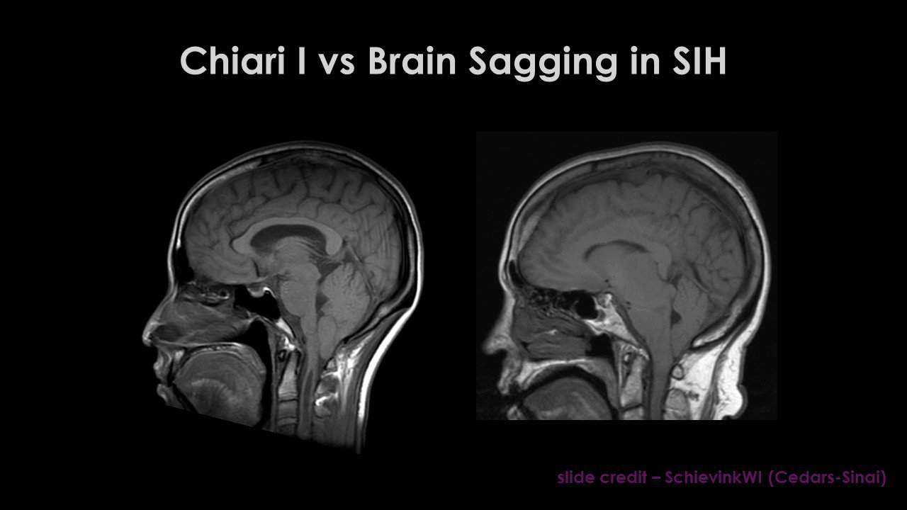
Neurologists review medical history thoroughly. Physical exams test balance and coordination. MRI scans reveal cerebellar tonsil descent clearly. Cine MRI shows abnormal cerebrospinal fluid flow. CT scans detect bone abnormalities accurately. Doctors measure tonsil extension beyond five millimeters. Associated syringomyelia appears as fluid cavities in the spine. Ultrasounds identify prenatal cases sometimes. X-rays check skull base structure. Comprehensive evaluation confirms the diagnosis definitively.

Non-Surgical Ways to Manage Chiari Malformation
Doctors prescribe pain medications for headaches effectively. NSAIDs reduce inflammation and discomfort quickly. Muscle relaxants ease neck tension successfully. Physical therapy strengthens core muscles gradually. Patients learn proper posture techniques daily. Massage therapy relieves muscle spasms regularly. Lifestyle changes avoid heavy lifting completely. Dehydration worsens symptoms dramatically. Patients stay hydrated consistently. Gentle exercises improve balance and coordination. Swimming provides low-impact activity benefits. Yoga enhances flexibility without strain. Monitoring with regular MRIs tracks progression closely. Asymptomatic patients avoid unnecessary interventions. Patients explore Chiari malformation treatments without surgery first.

Chiari Malformation Treatments: Surgical Options
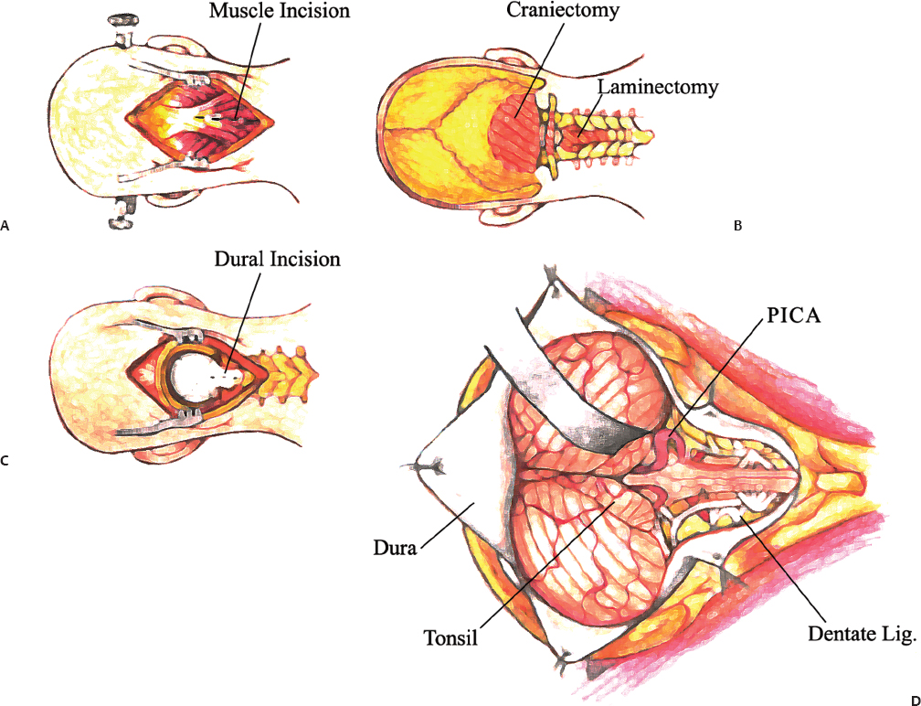
Surgeons perform posterior fossa decompression commonly. They remove a small section of occipital bone. This procedure creates extra space for the cerebellum. Pressure on the brainstem decreases immediately. Surgeons often remove the posterior arch of C1 vertebra. Duraplasty enlarges the dural sac with a patch. Cerebrospinal fluid flow restores naturally. Tonsillar shrinkage uses electrocautery precisely. Arachnoid dissection opens pathways for fluid. Laminectomy relieves spinal cord compression effectively. Surgeons address syringomyelia through decompression primarily. Shunts drain excess fluid in hydrocephalus cases. Chiari malformation treatments include advanced surgical techniques.
Additional Procedures Surgeons Use
Craniectomy removes bone to relieve intracranial pressure. Duraplasty patches expand the dura mater safely. Tonsil coagulation reduces herniated tissue volume. Intraoperative ultrasound guides precise bone removal. Endoscopic approaches minimize incision size effectively. Surgeons avoid obex plugging routinely. Individualized plans match patient anatomy perfectly. Pediatric cases favor bone-only decompression often. Adults benefit from duraplasty addition frequently. Reoperation rates remain low with proper technique.
Preparing for Chiari Malformation Surgery
Patients undergo preoperative MRI evaluations thoroughly. Blood tests check overall health status. Anesthesiologists assess surgical risks carefully. Surgeons explain expected outcomes clearly. Patients stop certain medications beforehand. Smoking cessation improves healing significantly. Nutrition supports immune function strongly. Family members learn postoperative care instructions. Mental preparation reduces anxiety effectively. Hospitals admit patients the day before surgery typically.
Recovery After Chiari Malformation Decompression
Patients stay in hospital three to seven days usually. Neck pain persists initially but improves gradually. Physical therapy begins early in recovery. Patients wear cervical collars sometimes. Gradual mobilization prevents complications effectively. Follow-up MRIs monitor cerebrospinal fluid flow. Symptoms resolve over several months often. Full recovery takes six to twelve months typically. Doctors schedule regular neurological exams. Pain medications taper down slowly. Patients resume light activities cautiously. Driving returns after clearance from surgeons. Doctors recommend Chiari malformation treatments based on symptoms.
Living Well with Chiari Malformation
Patients avoid contact sports and heavy lifting permanently. Straining during bowel movements triggers symptoms badly. Proper hydration prevents dehydration headaches. Stress management techniques reduce pain flares. Support groups connect patients worldwide. Adaptive equipment aids daily tasks effectively. Regular exercise maintains muscle strength. Balanced diet supports overall health. Patients track symptoms in journals consistently. Annual MRIs detect changes early. Quality of life improves dramatically with management. Successful Chiari malformation treatments lead to better quality of life.
Recent Advances in Chiari Malformation Care
Minimally invasive techniques reduce recovery time significantly. Intraoperative neuromonitoring prevents nerve damage. Genetic research identifies risk factors accurately. Advanced imaging improves diagnostic precision. Duraplasty materials enhance long-term outcomes. Pediatric protocols evolve continuously. Telemedicine enables remote follow-ups conveniently. Artificial intelligence analyzes MRI scans faster. Clinical trials test new decompression methods. Ongoing research improves Chiari malformation treatments steadily.
When to Seek Professional Help
Sudden severe headaches demand immediate attention. Worsening numbness requires urgent evaluation. Balance loss increases fall risks dangerously. Swallowing problems cause aspiration pneumonia. Vision changes signal pressure increases. Doctors evaluate new symptoms promptly. Early intervention prevents permanent damage. Emergency surgery addresses acute hydrocephalus. Patients contact specialists when symptoms interfere with life.
Final Thoughts on Chiari Malformation Management
Patients benefit from multidisciplinary care teams. Neurosurgeons, neurologists, and therapists collaborate effectively. Personalized plans address individual needs. Education empowers patients fully. Support networks provide emotional strength. Future therapies promise even better results. Awareness grows through advocacy efforts. Chiari malformation treatments offer promising outcomes for many.
READ ALSO: Exploring Skokie Sports Park Skokie IL: Your Ultimate Guide to Recreation and Fun