Appendicitis Ultrasound requires fast and accurate diagnosis. Doctors often rely on imaging to confirm inflammation. Ultrasound stands as a safe and effective option. It avoids radiation and delivers quick results. This guide explains how ultrasound helps detect appendicitis. It also covers preparation, accuracy, and benefits.
What Is Appendicitis?
Appendicitis occurs when the appendix becomes inflamed. The appendix is a small tube attached to the large intestine. Blockages often trigger inflammation. These blockages may include stool, infection, or swollen tissue. Symptoms usually appear suddenly and worsen quickly.:max_bytes(150000):strip_icc()/HDC-INFOGRAPHIC-Stages-of-Appendicitis-1-V1-1-b82cec7d8631478c8d2f84835557a9cc.png)
Common symptoms include:
- Severe pain in the lower right abdomen
- Nausea and vomiting
- Loss of appetite
- Fever
- Swelling in the abdomen
Early diagnosis prevents complications. Untreated cases may lead to rupture and infection.
Why Imaging Matters in Diagnosis
Doctors use physical exams and blood tests first. These methods suggest inflammation but lack certainty. Imaging confirms the diagnosis and reduces errors. It also helps avoid unnecessary surgery.
Among imaging options, appendicitis ultrasound plays a key role. It offers a quick and non-invasive solution. Doctors often choose it as the first-line test, especially for children and pregnant women.
How Ultrasound Works
Ultrasound uses high-frequency sound waves. These waves create images of internal organs. A handheld device called a transducer sends and receives signals. The machine converts signals into real-time images.
The process does not involve radiation. It makes ultrasound safe for repeated use. Technicians apply gel to improve contact and clarity.
Role of Ultrasound in Detecting Appendicitis
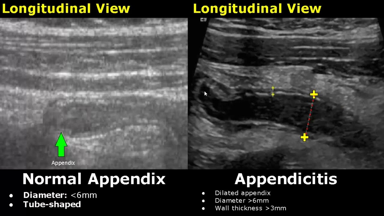
Appendicitis ultrasound helps visualize the appendix. A healthy appendix appears small and compressible. An inflamed appendix appears enlarged and rigid.
Key findings include:
- Enlarged appendix diameter
- Thickened walls
- Fluid collection around the appendix
- Increased blood flow
Doctors combine these findings with symptoms. This approach improves diagnostic accuracy.
When Doctors Recommend Ultrasound

Doctors recommend ultrasound when symptoms suggest appendicitis. It works best in early stages. It also helps rule out other conditions.
Situations where doctors prefer ultrasound include:
- Children with abdominal pain
- Pregnant women
- Patients avoiding radiation exposure
- Initial evaluation before CT scans
In many cases, appendicitis ultrasound provides enough evidence for diagnosis.
Preparing for the Procedure
Preparation improves image quality. Patients may need to fast for several hours. A full bladder sometimes helps in better visualization.
Follow these steps:
- Avoid eating for 6–8 hours
- Drink water if instructed
- Wear loose clothing
- Inform the technician about symptoms
Proper preparation ensures clearer results.
Step-by-Step Procedure
The procedure remains simple and painless. It usually takes 20 to 30 minutes.
Here is what happens:
- The patient lies on an examination table
- The technician applies gel on the abdomen
- The transducer moves across the lower abdomen
- The technician applies gentle pressure
- Images appear on the monitor
Patients may feel slight discomfort during pressure. This helps locate the inflamed appendix.
Accuracy of Ultrasound in Diagnosis
Accuracy depends on several factors. These include technician skill and patient body type. Ultrasound performs best in thin patients and children.
Studies show high accuracy rates when experts perform the scan. However, results may vary in adults with higher body fat.
Despite limitations, appendicitis ultrasound remains reliable. It often serves as the first diagnostic step.
Advantages of Ultrasound
Ultrasound offers many benefits compared to other imaging methods.
Safe and Radiation-Free
Ultrasound does not use radiation. This makes it safe for all age groups. Pregnant women benefit the most from this feature.
Quick and Accessible
Hospitals and clinics widely offer ultrasound services. The procedure provides immediate results.
Cost-Effective
Ultrasound costs less than CT scans. It reduces healthcare expenses for patients.
Real-Time Imaging
Doctors observe movement and blood flow instantly. This improves diagnostic confidence.
Limitations of Ultrasound
Ultrasound also has some drawbacks. Understanding these helps set realistic expectations.
Operator Dependency
Results depend on the technician’s experience. Skilled professionals produce better images.
Limited Visibility
Gas in the intestines may block clear images. Obesity also reduces accuracy.
Inconclusive Results
Sometimes ultrasound cannot confirm appendicitis. Doctors may order further imaging in such cases.
Even with limitations, appendicitis ultrasound remains a valuable tool.
Comparing Ultrasound with CT Scan
Doctors often compare ultrasound with CT scans. Each method has its own strengths.
Ultrasound
- No radiation
- Lower cost
- Ideal for children and pregnant women
- Quick results
CT Scan
- Higher accuracy in adults
- Better visualization of complications
- More detailed images
Doctors usually start with ultrasound. They move to CT if results remain unclear.
Signs of Complicated Appendicitis
Ultrasound can also detect complications. These require urgent medical attention.
Signs include:
- Abscess formation
- Perforation of the appendix
- Fluid collection in the abdomen
Early detection prevents serious outcomes. It also guides treatment decisions.
Role in Pediatric Diagnosis
Children often struggle to describe pain. This makes diagnosis challenging. Ultrasound helps reduce uncertainty.
Doctors prefer appendicitis ultrasound for children. It avoids radiation exposure and provides quick answers. Pediatric specialists rely on it as the first step.
Role During Pregnancy
Pregnancy limits imaging options. Radiation-based tests pose risks to the fetus. Ultrasound becomes the safest choice.
Doctors use it to evaluate abdominal pain in pregnant women. It helps differentiate appendicitis from other conditions.
After the Diagnosis
Doctors decide treatment after confirming appendicitis. Most cases require surgery. Surgeons remove the inflamed appendix.
In some cases, antibiotics may help. Doctors monitor these patients closely. Early diagnosis improves recovery outcomes.
Recovery and Follow-Up
Recovery depends on the severity of the condition. Simple cases heal quickly after surgery. Complicated cases may take longer.
Doctors may use ultrasound during follow-up. It helps check for infection or fluid buildup.
Patients should follow medical advice carefully. Proper care ensures faster healing.
Tips for Better Diagnostic Results
Patients can improve accuracy by following simple tips:
- Seek medical help early
- Describe symptoms clearly
- Follow preparation instructions
- Choose experienced medical facilities
These steps enhance the effectiveness of appendicitis ultrasound.
Future of Ultrasound in Appendicitis Diagnosis
Medical technology continues to evolve. Advanced ultrasound machines offer better image quality. Artificial intelligence may improve interpretation accuracy.
Portable ultrasound devices are also emerging. These allow quick diagnosis in emergency settings.
The future looks promising for faster and more precise detection.
Conclusion
Appendicitis requires timely diagnosis and treatment. Ultrasound provides a safe and efficient solution. It helps doctors confirm inflammation without radiation risks. Appendicitis ultrasound remains a first-line diagnostic tool. It works well for children, pregnant women, and early cases. While limitations exist, its benefits outweigh the drawbacks.
Understanding the procedure helps patients feel more confident. Early diagnosis saves lives and prevents complications. Always seek medical attention if symptoms appear.
READ ALSO: Can Antibiotics Make You Constipated? Understanding the Gut Connection and Effective Solutions